
주문제작 서비스 문의
찾으시는 제품이 저희 사이트에 없으신가요? 단백질 주문 제작 서비스를 이용해보세요!
주문제작 서비스 문의 >>


 Limited Edition Golden Llama is here! Check out how you can get one.
  Limited Edition Golden Llama is here! Check out how you can get one.
Offering SPR-BLI Services - Proteins provided for free! Offering SPR-BLI Services - Proteins provided for free!
Here come GMP Grade Cytokines!Free Sample is available! Here come GMP Grade Cytokines!Free Sample is available!
> Immunohistochemistry (IHC) Antibody
 Antibody")
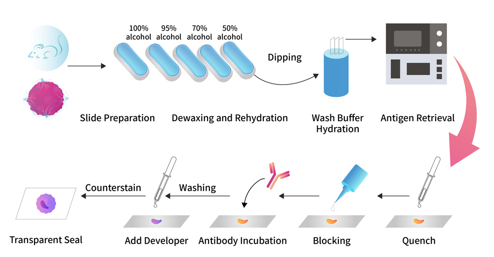
ACROBiosystems launched the antibody suitable for immunohistochemistry verified by ACRODiagnostic medical pathology platform, with sufficient methodological verification and kit performance verification data to support histochemical verification and development of multiple applications as immunohistochemical kits, etc.!


| Cat. No. | Molecular | Product Description | Preorder/Order |
|---|
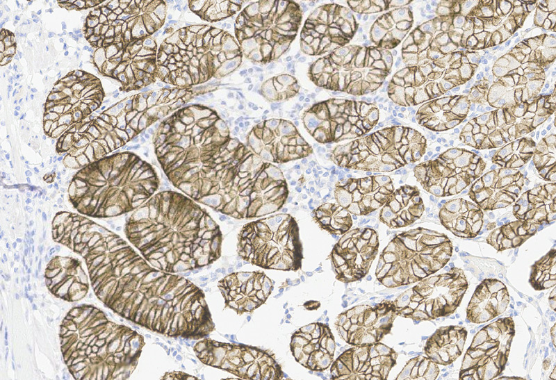
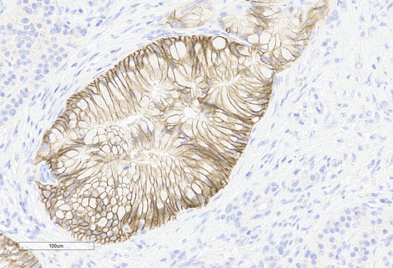
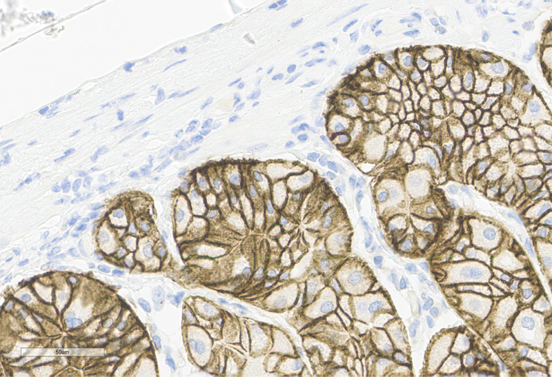
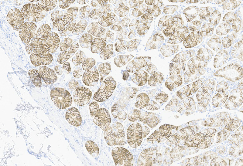
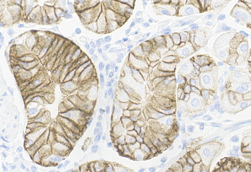
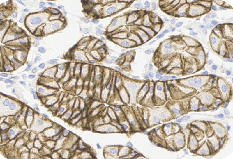
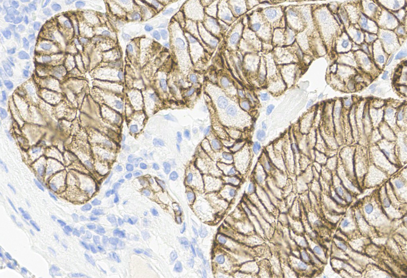
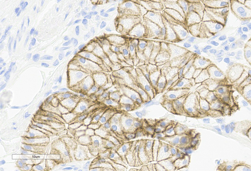
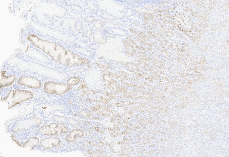
Human Stomach Tissue, 4X
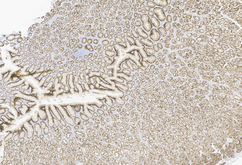
Human Stomach Tissue, 20X
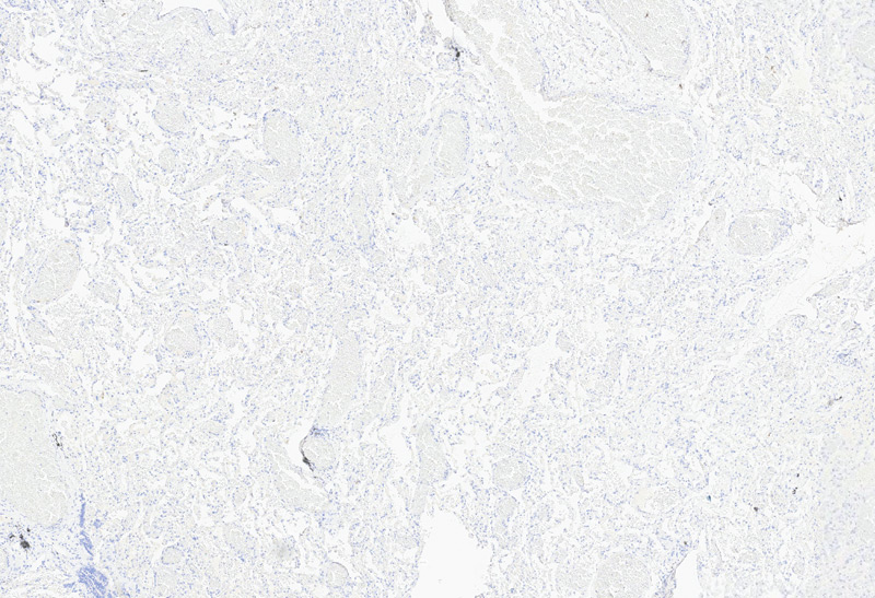
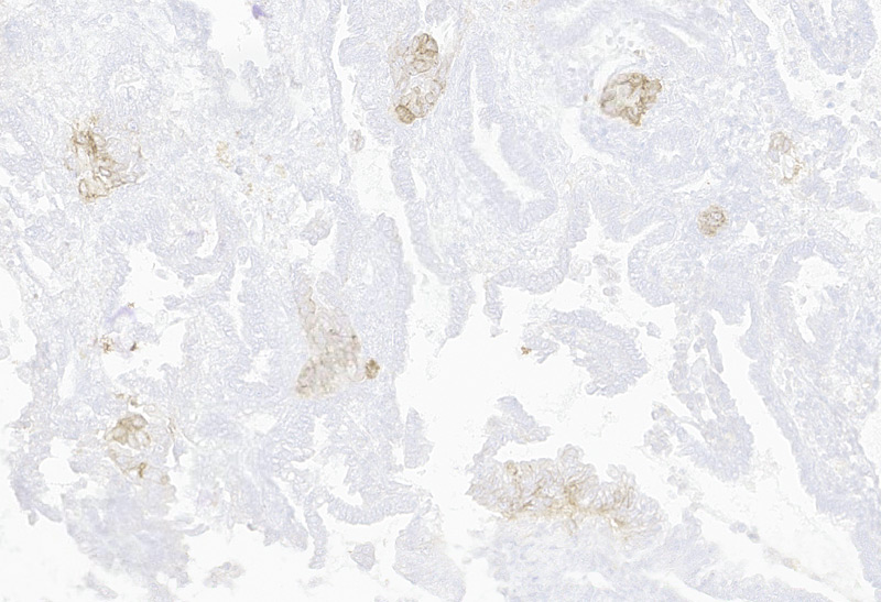
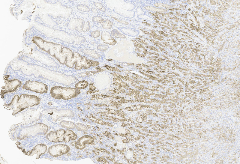
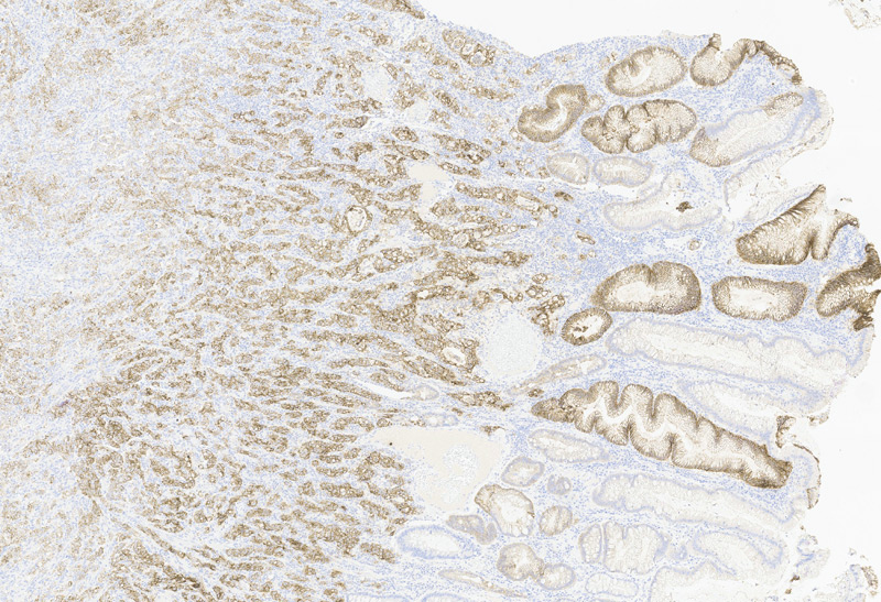
Human Lung Tissue, 4X
Human Lung Tissue, 20X
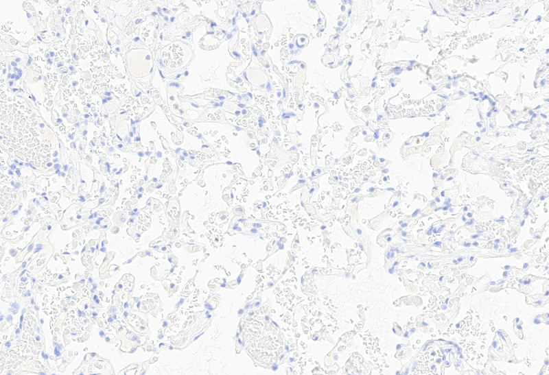
* The human gene Claudin-18 has two protein isoforms, Claudin-18.1 and Claudin-18.2, which differ within the N-terminal 69 amino acids. Claudin-18.1 expression is restricted to the lungs, whereas Claudin-18.2 expression is restricted to the stomach.
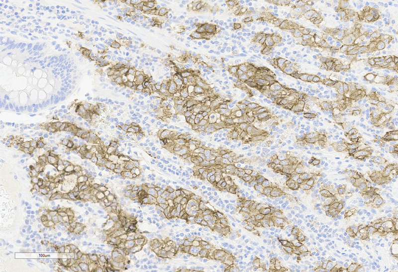
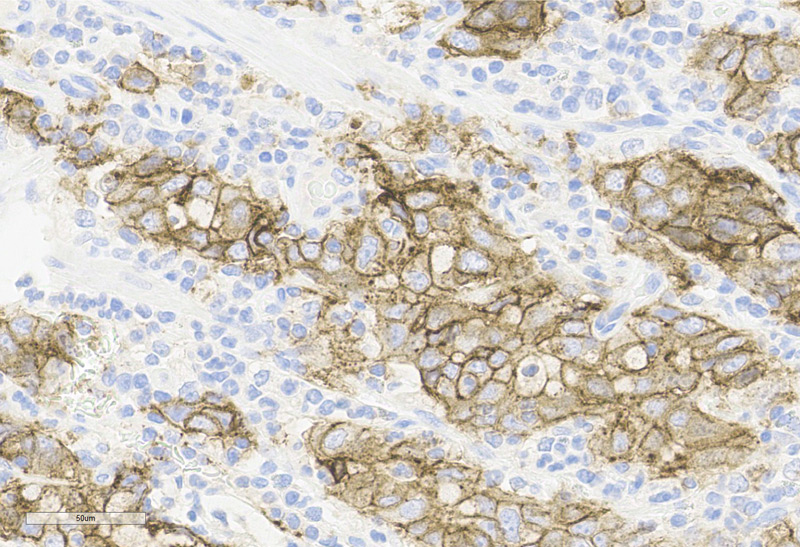
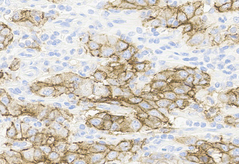
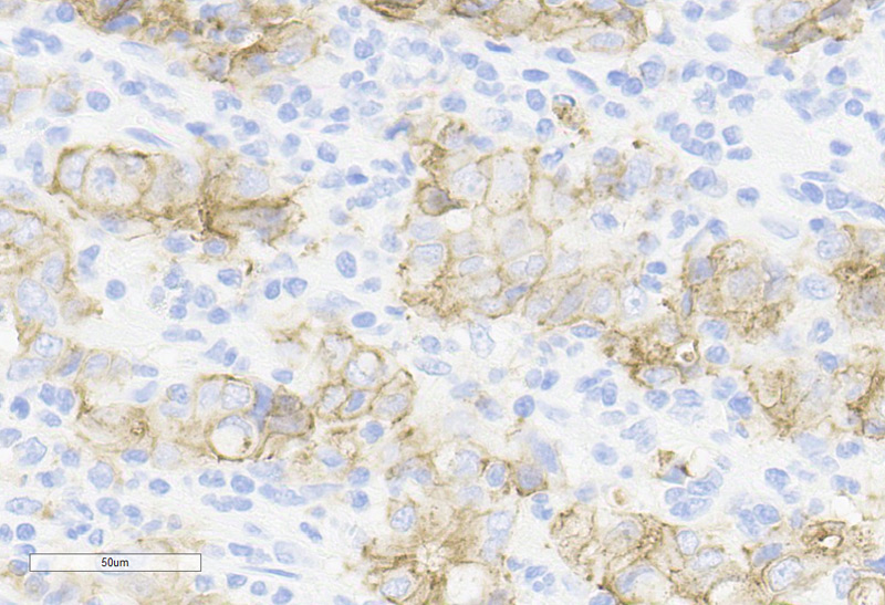
Human Gastric Cancer, 20X
Human Colorectal Cancer, 20X
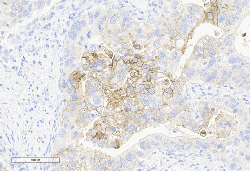
Human Pancreatic Cancer, 20X
Human Ovarian Cancer, 20X
Leica BOND-III-10X
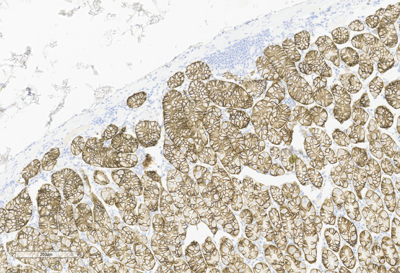
Leica BOND-III-40X
Dako link 48-10X
Dako link 48-40X
Claudin18.2 (ACRO)
1:1000-40X
Claudin18.2 (Competitor A)
1:200-40X
Claudin18.2 (Competitor A)
1:500-40X
Claudin18.2 (ACRO)
1:1000-4X
Claudin18.2 (Competitor A)
1:200-4X
Claudin18.2 (Competitor A)
1:500-4X
Claudin18.2 (ACRO)
1:1000-40X
Claudin18.2 (Competitor A)
1:200-40X
Claudin18.2 (Competitor A)
1:500-40X

This web search service is supported by Google Inc.


 A-Z
A-Z


